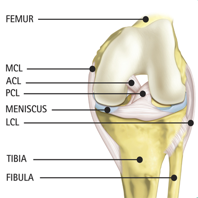
Healthy Knee Anatomy
The knee is a hinge joint formed by the tibia (shin bone), femur (thigh bone) and patella (knee cap). The joint surface where these bones meet is covered with articular cartilage, a smooth substance that enables the knee joint to move fluently. There are also two wedge shaped pieces of tough, rubbery cartilage called meniscus, which sits between the thigh and leg bones and acts as a shock absorber. All remaining surfaces of the knee are covered by a thin, smooth tissue liner called the synovial membrane, which lubricates the knee and reduces friction to nearly zero. The patella, with ligaments, is essential for transferring the strength of the thigh muscle to the muscles of the lower leg. Ligaments are tough bands of tissue that connect ends of bones together. The cruciate and collateral ligaments provide the knee with stability. Two important ligaments are found on either side of the knee: medial collateral ligament (MCL) and lateral collateral ligament (LCL) which prevent excessive side-to-side movement. The anterior cruciate ligament (ACL) and posterior cruciate ligament (PCL) are found behind and in front of the knee and control front-to-back movements. Tendons attach muscles to bones. The largest tendon around the knee is the patellar tendon, which connects the patella to the tibia, covers the patella, and continues up the thigh. As the patellar tendon continues up the thigh, it is referred to as the quadriceps tendon since it attaches to the quadriceps muscles.
Movement of the Knee Joint
The extensor mechanism is the motor that drives the knee joint and allows us to walk. It sits in front of the knee joint and is made up of the patella, patellar tendon, quadriceps tendon and quadriceps muscles. When the quadriceps muscles contract, the knee joint straightens. The hamstring muscles are located in the back of the knee and thigh. When these muscles contract, the knee bends.